Language is a unique and essential function of human beings and is critical to quality of life. For patients with a brain tumor near putative language cortex, neurosurgeons may use functional magnetic resonance imaging (fMRI) presurgical language mapping to assess the risks of surgery-induced permanent language deficits against the potential benefits of maximal resection. Critical barriers to clinical deployment of presurgical fMRI are that
- The validity is contingent on the patient’s ability to perform precisely timed phonological and semantic tasks. However, in our center, up to 50% of patients assigned to fMRI have a language deficit that may affect task performance and invalidate the mapping.
- Expertise in administering language tasks is insufficient in many clinical settings.
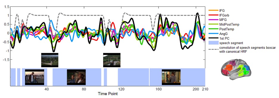
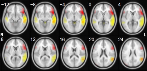
To tackle these challenges, we have investigated and demonstrated the feasibility and effectiveness of task-free, e.g., resting-state and movie-watching, fMRI paradigms in mapping language areas in individual healthy subjects and neurosurgical patients.
Resting-state fMRI
Movie-watching fMRI

