Intracranial Electrical Stimulation Testing (ECS) and Intracranial EEG
Intracranial electrophysiologic studies in awake people can provide a unique window into the functioning of the human brain. This type of study is performed in certain patients undergoing brain surgery in order to minimize the risks of postoperative neurologic dysfunction. If patients agree and give informed consent it is also possible to collect data from these patients which can help answer many basic neuroscientific questions.
Advantages of Intracranial EEG
iEEG methods are well suited to the study of task-related neuronal activity. The problems of source localization that pose serious challenges to EEG and even MEG analyses are far less acute when electrical recordings are taken from intracranial electrodes (either electrode grids placed directly on the cortical surface or implanted depth electrodes). These electrodes sample from much smaller volumes (Nunez, Srinivasan et al. 1997) , are not subject to distortions introduced by the skull and scalp (Pfurtscheller and Cooper 1975) and are relatively impervious to artifacts produced by eye movements and blinks. These factors contribute to the high signal-to-noise ratio characteristic of iEEG recordings, which, in turn, help locate the areas of steep voltage gradients indicative of signal sources in the human brain (Kahana, Seelig et al. 2001).
Awake Surgery, Cognitive Testing, and Intracranial Electrodes
Neurosurgical resection is the primary therapeutic intervention in the treatment of brain tumors and medically refractory epilepsy. In both cases, the challenge is to completely remove the affected tissue while sparing eloquent brain tissue which may be immediately adjacent. These resections may be difficult because the visual distinction between diseased tissue and adjacent normal brain may be subtle or impossible. The addition of awake surgery under local anesthesia allows monitoring of the neurological integrity of the patient. During awake surgery, electrical stimulation of the cortical surface, through a handpiece or cortical grid, is used to locate motor, sensory and language areas, with muscle contractions, paresthesias, or speech arrest respectively. High frequency cortical stimulation, despite its name, actually disrupts brain function clinically. Electrocorticography may also be used to identify regions of epileptiform activity. Another approach used frequently in epilepsy surgery uses surgically implanted electrodes with chronic (~ 1 week) extra-operative monitoring and testing. These techniques are essential to the safe and effective treatment of these patients. They also provide a unique opportunity to study the human brain in vivo.
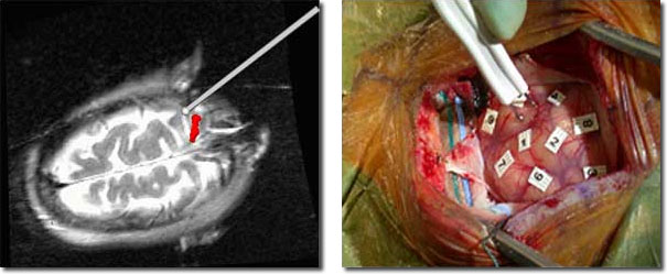
Intraoperative use at BWH of fMRI and ECS brain mapping for localization of eloquent cortex. (left) Intra-operative imaging
demonstrates coregistration of preoperative fMRI motor mapping data (red) with updated MR image of brain and probe position
(right) Intra-operative photograph showing electrical cortical stimulation mapping with markers indicating areas which have been tested.
The Golby lab is collaborating with the Computational Memory Lab at the University of Pennsylvania and Joseph Madsen at Children’s Hospital to use iEEG to study memory processing. Over 25 patients performing a variety of memory tasks have been successfully studied. Data has been acquired from a large number of behavioral paradigms and from many brain areas (see figure below).
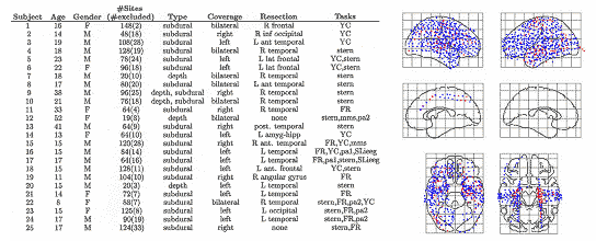
(left) Table summarizing all subjects studied to date by the mentor’s laboratory. (YC= yellow cab the virtual maze task;
stern=classic Sternberg variant; FR=free recall variant; pa1&2=paired associate variants; mms=multi modal Sternberg
(pictures, letters, words, spatial) (right) Schematic of all recording sites from all subjects across all experiments in the
mentor’s lab which have been included and excluded from analyses. Electrode locations are represented as filled
(blue-included; red-excluded) circles on a standardized brain. Top row shows the lateral surfaces of the brain; middle
row shows the medial surfaces; and the bottom row shows the dorsal and ventral surfaces.