A Collaboration with: Michael Kahana at Brandeis University
This project uses both intracranial electroencephalography (iEEG) and functional Magnetic Resonance Imaging (fMRI) to study the organization of memory processing in the human brain. These techniques will be applied in patients undergoing surgical evaluation and treatment for brain tumors and epilepsy. These experiements aim to:
- shed light on the organization of memory processing in the human brain,
- refine these tools so that they may aid in the determination of individual patient anatomy, and improve surgical planning, and
- promote a better understanding of the relationship between the task-induced fMRI activation based on the hemodynamic blood oxygen level dependent (BOLD) signal and task-induced changes in neural electrical activity.
MR- and EEG-based techniques possess complementary strengths and weaknesses in terms of spatial and temporal resolution, vulnerability to artifact, and brain coverage. Combining these two modalities can reveal information about of the functional architecture of cognition that would be absent from either method used in isolation. We are using both passive recording from the brain as well as attempted task disruption to understand better the relationship between activation and task performance.

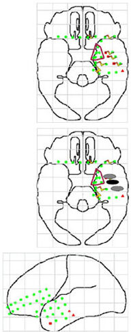
Brain maps from a study of attempted task disruption in a patient showing electrode locations (in green). Sites marked in red are electrodes that showed correlations between oscillatory activity and the subsequent memory effect (squares positive correlation; triangles negative correlation). Lightning bolts connect sites of bipolar stimulation. The ellipses show sites where stimulation during encoding resulted in poor memory for the items (grey, 1/3 items remembered; black, 0/3).