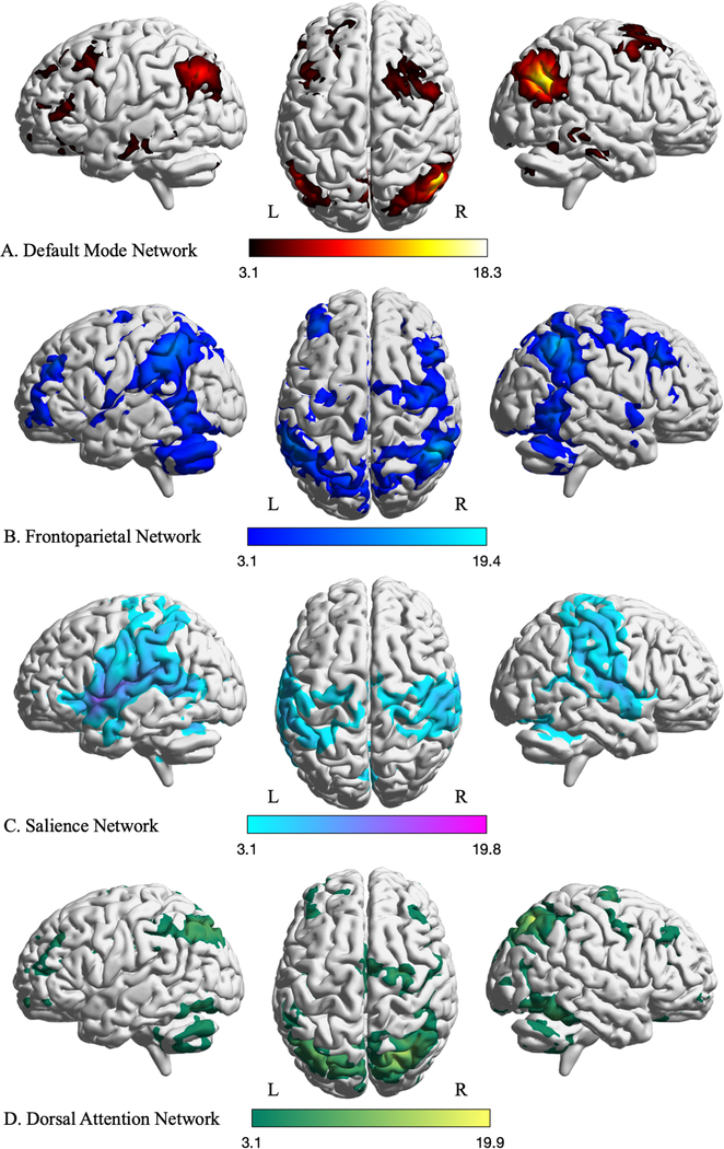
Presurgical brain mapping with fMRI has focused on the identification and preservation of so-called “eloquent” brain functions (i.e., motor and language), however, mapping of “non-eloquent” areas, such as cognitive and emotional networks, is essential for preserving and improving patients’ quality of life. Resting-state fMRI can be used to simultaneously explore multiple resting-state networks (e.g., default mode, attention, salience, and executive functional networks) related to higher order brain functions.
In addition to resting-state fMRI, an emerging fMRI paradigm with naturalistic stimuli, i.e., movie clips, can be used to map emotion networks.
